Often called “The Silent Thief of Sight”, glaucoma is a group of disorders that damage the optic nerve, causes vision loss if left untreated. Glaucoma is one of the leading causes of blindness. Glaucoma symptoms typically develop slowly and by the time there is noticeable vision loss, the condition is late-stage. Early detection is key to treating and preventing permanent vision loss.
How To Test For Glaucoma
Early detection is the best way to prevent vision loss due to glaucoma and that starts with having a comprehensive eye exam every year. Glaucoma cannot be diagnosed with one single test. Your doctor must look at different pieces of the puzzle to make that determination. A glaucoma evaluation will normally consist of all or some of these tests:
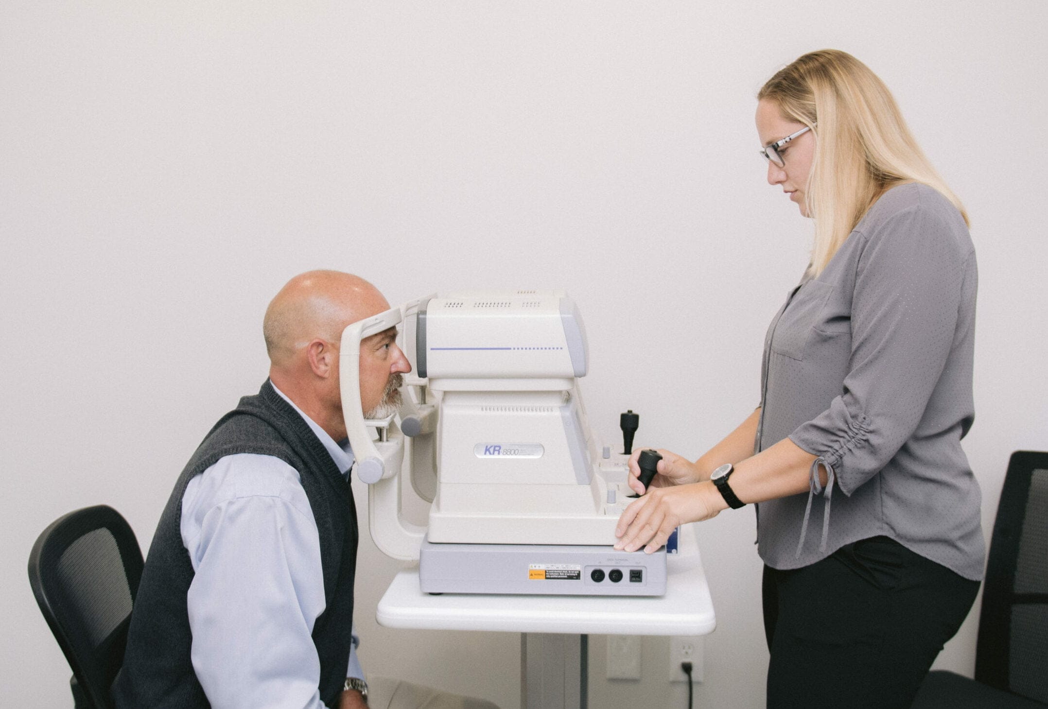
[media id=”37654″ alt=”patient sitting in front of an air puff tonometer during an eye exam”]
Eye Pressure Test (Tonometry)
Increased eye pressure can be a sign of glaucoma. A common way to check eye pressure is the “air puff test”, a method used to measure the pressure inside your eye without touching your eye. Another name for it is non-contact tonometry. The quick air puff gives your eye doctor an eye pressure reading known as intraocular pressure (IOP). There are other ways to measure IOP that involve using a probe on the eye and your doctor will often do this in the exam room if your IOP was abnormal. If your IOP is too high, your eyes are either producing too much fluid or not draining enough fluid. Eye drops can help keep IOP at an acceptable level. If those fail, surgery might be necessary.
Dilated Eye Exam
Eye drops are used to dilate the pupil so that the doctor can see through your eye to get a better view of your optic nerve, blood vessels, and retinal tissue. This helps your doctor assess the shape, color, depth, size, and vessels of the optic nerve to determine your risk for glaucoma.
Optomap or OPTOS
An Optomap is another tool that we have in select locations that captures a wide-field image of the eye, similar to what your doctor sees during a dilated assessment. The image allows the doctor to view the retina in more detail, diagnose diseases quickly, and create a permanent record that can be used to compare side by side in future exams. The Optomap is a valuable tool, but dilation may still be necessary depending on the patient and condition.
Visual Field Test (Perimetry)
A common symptom of glaucoma is peripheral vision loss; a visual field test is a simple way to test for subtle loss of field in one eye that, as a patient, you can’t detect on your own. During this test, small flashes of light will be presented in your peripheral vision. Glaucomatous vision loss tends to present in distinct patterns that correspond to nerve fiber loss in the optic nerve itself. This is a subjective test (relies on the patient to respond) that can be compared to some of the objective tests (no response from the patient) to determine if damage to the nerve has occurred or if your glaucoma has gotten worse.
Angle Test (Gonioscopy)
The doctor will use a mirrored lens called a gonioscopy lens to touch the numbed cornea. The angle test is simple, quick, and does not hurt. This helps determine whether the angle is open and wide or narrow and closed. The “angle” is the area where the cornea (the clear part in the front of the eye) meets the iris (the colored part of the eye). The majority of the fluid that is created inside the eye must exit the eye by passing through the meshwork of the angle. If it’s blocked or narrow, this can cause fluid to build up and eye pressure to increase, and possible damage to the nerve.
Corneal Thickness Test (Pachymetry)
While your eye is numbed, the corneal thickness test painlessly measures the thickness of the cornea with a small probe. The thickness measurement provides crucial information to your doctor to better understand your IOP and the overall health of your eye. If your corneal is thicker than average your IOP measurement may read falsely higher and if it’s thinner than average it may read falsely lower than average.
TYPES OF GLAUCOMA
- Primary open-angle glaucoma, which is most common, will develop over time and not show any symptoms initially.
- Angle-closure glaucoma, which is less common, but is a medical emergency that can cause vision loss within a day of onset. Patients are very symptomatic with eye pain, blur, redness, or nausea.
- Secondary glaucoma occurs as a result of an injury or other eye diseases.
- Normal-tension glaucoma, which causes optic nerve damage even though the eye pressure remains in the “normal” range.
[media id=”37591″ alt=”patient asking optometrist questions about glaucoma”]
ARE YOU AT RISK?
Everyone should get have an eye exam annually, but if you meet one or more of the criteria below, be sure to take extra care when talking to your doctor about the risks for glaucoma.
- Being over 60. If you are older than 60, you are six times more likely to have glaucoma.
- Family history. Glaucoma runs in the family, increasing chances by four to nine times.
- Steroid medication use. One study found that heavy use of inhaled steroids for asthma boosted the risk by 40 percent.
- Ethnicity. African-Americans and Hispanics are six to eight times more likely to develop glaucoma than Caucasians.
- Eye injury. Even if it is only a black eye, eye injuries can cause glaucoma years after impact. Use protective eyewear for activities that may cause eye injury, such as sports like boxing or baseball or using power tools.
Is There A Cure for Glaucoma?
There is no cure for glaucoma. However, treatment and regular eye exams can help slow or prevent vision loss, especially if you catch the disease in its early stages. Glaucoma symptoms aren’t immediately noticeable. Until there is a cure, prevention starts with an annual comprehensive eye exam at Dr. Tavel.

